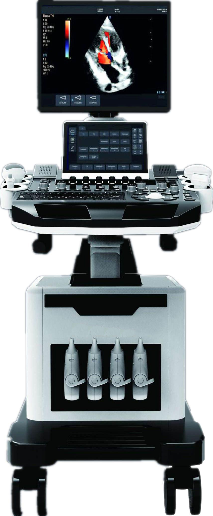
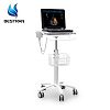
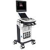
Trolley color doppler Ultrasound Diagnostic System
Trolley color doppler Ultrasound Diagnostic System
|
1: |
Summary of main specifications and system of cart type color Doppler ultrasound |
|
1.1 |
Trolley type all digital color Doppler ultrasonic mainframe |
|
1.2 |
Ultrasonic host operating system: Windows operating system |
|
1.3 |
Applications: Abdomen, obstetrics, gynecology, heart, urinary system, small organs, superficial, blood vessels, pediatrics, newborns, musculoskeletal |
|
1.4 |
Probes: Convex probe, Tran-vaginal probe, Linear probe, Micro-convex probe, Cardiac probe,4D Volume probe |
|
1.5 |
Applications and report: Abdominal,OB,GYN,Cardiac,Urinary,Small Parts,Superficial, Vascular, Pediatrics, Advanced measurement software packages, report software packages, case management software packages, etc. |
|
☆1.6 |
carotid artery intima measurement thickness(IMT) |
|
☆1.7 |
Automatic spectral envelope measurement |
|
1.8 |
Full digital transmission and reception of beam synthesizer |
|
1.9 |
Color Doppler imaging(C) |
|
1.10 |
Pulse Doppler Imaging(PW) |
|
☆1.12 |
Coherent Contrast imaging(CCI) |
|
1.12 |
Continuous wave Doppler imaging(CW) |
|
☆1.13 |
B/C/D Real-time three synchronous imaging |
|
☆1.14 |
Power Doppler imaging(PDI) |
|
☆1.15 |
Direct power Doppler imaging(DPDI) |
|
1.16 |
M mode imaging |
|
☆1.17 |
Anatomic M mode imaging |
|
☆1.18 |
Color Doppler M mode imaging |
|
☆1.19 |
Elastography |
|
☆1.20 |
Tissue Doppler imaging(TDI) |
|
☆1.21 |
Strain rate imaging (SRI) |
|
1.22 |
Tissue harmonic imaging(THI) |
|
1.23 |
Fusion harmonic imaging(FHI) |
|
1.24 |
Speckle Reduce imaging(SRI) |
|
☆1.25 |
Panoramic imaging |
|
☆1.26 |
Deflection imaging |
|
☆1.27 |
Trapezoidal imaging |
|
1.28 |
Adaptive velocity optimization |
|
☆1.29 |
Free hand 3D |
|
1.30 |
Real time 3D imaging(3D/4D) |
|
1.31 |
DICOM3.0 |
|
1.32 |
Monitor:≥19 inch,high definition ultrasonic display |
|
1.33 |
≥10 inch touch screen |
|
1.34 |
Physical clipboard: save the image on the left side of the screen, which can be directly saved or deleted. |
|
1.35 |
The system has the function of on-the-spot upgrade |
|
1.36 |
Presupposition: for different inspection of the viscera, preset the inspection conditions for the best image, reduce the adjustment of the operation, and the commonly used external adjustment and combination regulation. |
|
1.37 |
Probe interface: 4 |
|
1.38 |
Chinese and English System, Chinese and English input, optional |
|
1.39 |
Depth:≥360mm; |
|
1.40 |
Extended imaging |
|
2: |
Probes |
|
2.1 Convex probe |
Fundamental Frequency:2.0MHz/2.3MHz/2.5MHz/3.0MHz/3.5MHz/4.0MHz/4.6MHz/5.0MHz/5.4MHz, Harmonic Frequency: 4.0MHz/4.6MHz/5.0MHz, |
|
2.2 Linear probe |
Fundamental Frequency:4.0MHz/4.6MHz/5.0MHz/6.0MHz/7.0MHz/8.0MHz/9.2MHz/10.0MHz/12.0MHz/13.3MHz, Harmonic Frequency: 8.0MHz/9.2MHz/10.0MHz, |
|
2.3 Trans-vaginal probe |
Fundamental Frequency: 3.0MHz/3.5MHz/4.0MHz/5.0MHz/5.4MHz/6.0MHz/7.0MHz/8.0MHz/10.0MHz, Harmonic Frequency: 6.0MHz/7.0MHz/8.0MHz, |
|
2.4 Micro-convex probe |
Fundamental Frequency: 3.0MHz/3.5MHz/4.0MHz/5.0MHz/5.4MHz/6.0MHz/7.0MHz/8.0MHz, Harmonic Frequency: 6.0MHz/7.0MHz/8.0MHz, |
|
2.5 Cardiac probe |
Fundamental Frequency:1.7MHz/1.9MHz/2.1MHz/2.5MHz/3.0MHz/3.4MHz/3.8MHz/4.2MHz/5.0MHz, Harmonic Frequency: 3.4MHz/3.8MHz/4.2MHz, |
|
2.6 4D Volume probe |
Fundamental Frequency: 2.0MHz/2.5MHz/3.0MHz/3.3MHz/3.7MHz/4.0MHz/5.0MHz/6.0MHz, Harmonic Frequency: 4.0MHz/5.0MHz/6.0MHz, |
|
3: |
2D imaging mode |
|
3.1 |
Gain:0-100,Step 2 adjustable |
|
3.2 |
TGC:8 segment adjustable |
|
3.3 |
Maximum focus point:≥7, which can be moved throughout the whole process. |
|
3.4 |
Speckle reduction:0-5,5 level |
|
3.5 |
Space Synthesis:0-2,2 level(Liner probe: 3 level, cardiac probe:0) |
|
3.6 |
Dynamic:30-180,35 level,step 5 adjustable |
|
3.7 |
Line density:low、middle、high,3 level |
|
3.8 |
Frame correlation:0-4,4 level |
|
3.9 |
Noise reduction:0-5,5 level |
|
3.10 |
Edge Enhancement:0-5,5 level |
|
3.11 |
Sound power:2-10, 9 level |
|
3.12 |
Grey scale:0-67, 67 level |
|
3.13 |
False color:0-67,67 level |
|
3.14 |
Image style:Soft-Comparison,2 level |
|
|
The screen has real-time display of voice power, probe frequency, dynamic range, pseudo color, gray scale and other 11 parameters can be adjusted |
|
4: |
Color Doppler imaging mode |
|
4.1 |
Blood gain:0-100,Step 2 |
|
4.2 |
Parameter display:Velocity、Variance |
|
4.3 |
B-Restrain(B/W restrain):0-7, 7 level |
|
4.4 |
Speed Through:0-8, 8 level |
|
4.5 |
Sampling number:6-24, 7 level
|


