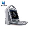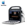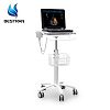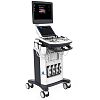


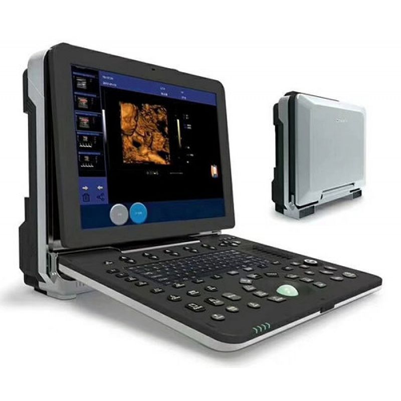
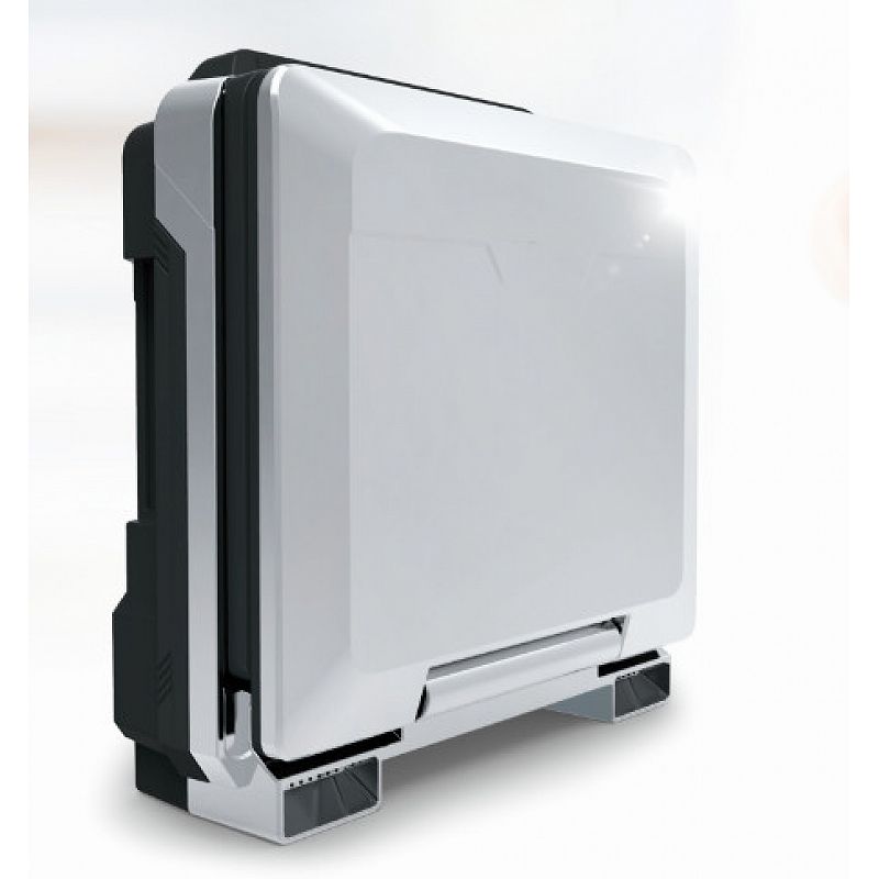
4D Portable color doppler machine
|
MODEL |
BT-UD300 3.0 VERSION
|
|
Computer specs |
Windows Embedded operation system (EN language) 15’’ medical monitor(1280*1024) Intel i5 processor 4G RAM 120G SSD(standard)+500G HDD(optional) |
|
Imaging Modes |
2D, 3D, 4D, Color/PW/CW/Power/Directional Color Power Doppler, |
|
Features |
Compound Imaging, iBank database |
|
DICOM Modes |
Store, Print, Working list, Storage Commitment, Structured Reports |
|
Export Options |
DICOM, Ethernet, JPG/BMP/PNG,AVI, |
|
Input/Output |
VGA, 2 USB Ports, Ethernet, Dicom, Built-in speakers |
|
Transducer Types |
Convex, Linear, Sector Phased, Micro Convex, |
|
Applications |
Abdominal, OB/GYN, Urology, Cardiac, Vascular, |
|
Probe Ports |
2 Active |
|
Cine Memory |
>10 seconds, 750frames |
Probe Specifications
Convex probe- Adult abdomen application
Frequency: 2.5, 3, 3.5, 4, H4, H5MHz
Power:5-100% (arithmetic progression of 5: 5,10,15...100)
Gain:0-100
Dynamic range: 20-280% (geometric progression of 2 start from 20: 20,40,60...280)
Gray map:0-7
Frame correlation:0-4
Filtering:0-4
Image denoising:0-14
Scanning depth:3-27.3cm
Body mark:17
Scanning range:50-100% (arithmetic progression of 10 start from 50: 50,60,70...100)
Focus point:6
Pseudo color map :0-11
Linear density:64,128,256
TSI: normal, fat, fluid, muscle
Reversal:up/down, left/right
Compound frequency: on/off
Automatic optimization: on/off
Space compound: on/off
Linear probe-Carotid application
Frequency: 6, 7.5, 8.5, 10, H10 MHz
Power:5-100% (arithmetic progression of 5: 5,10,15...100)
Gain:0-100
Dynamic range: 20-280% (geometric progression of 2 start from 20: 20,40,60...280)
Gray map:0-7
Frame correlation:0-4
Filtering:0-4
Image denoising:0-14
Scanning depth:2-11cm
Body mark:13
Scanning range:50-100% (arithmetic progression of 10 start from 50: 50,60,70...100)
Focus point:5
Pseudo color map :0-11
Linear density:64,128,256
TSI: normal, fat, fluid, muscle
Reversal:up/down, left/right
Steering: left/right
Trapezoid Imaging: on/off
Compound frequency: on/off
Automatic optimization: on/off
Space compound: on/off
Cardiac probe- Adult cardiac application
Frequency: 2.5, 3, 3.5, 4, H3, H4 MHz
Power:5-100% (arithmetic progression of 5: 5,10,15...100)
Gain:0-100
Dynamic range: 20-280% (geometric progression of 2 start from 20: 20,40,60...280)
Gray map:0-7
Frame correlation:0-4
Filtering:0-4
Image denoising:0-14
Scanning depth:3-27.3cm
Body mark:7
Scanning range:50-100% (arithmetic progression of 10 start from 50: 50,60,70...100)
Focus point: 5
Pseudo color map :0-11
Linear density:64,128,256
TSI: normal, fat, fluid, muscle
Reversal:up/down, left/right
Compound frequency: on/off
Automatic optimization: on/off
Space compound: on/off
4D probe R40- Obstetrics applications
Central frequency: H5.0MHz
Multi-frequency: 3.0, H5.0, 6.0, 4.5, 3.0, 2.0MHz
Power:5-100% (arithmetic progression of 5: 5,10,15...100)
Gain:0-100
Dynamic range: 20-280% (geometric progression of 2 start from 20: 20,40,60...280)
Gray map:0-7
Frame correlation:0-4
Filtering:0-4
Image denoising:0-14
Scanning depth:3-27.3cm
Body mark:7
Scanning range:50-100% (arithmetic progression of 10 start from 50: 50,60,70...100)
Focus point: 6
Pseudo color map :0-11
Linear density:64,128,256
TSI: normal, fat, fluid, muscle
Reversal:up/down, left/right
Compound frequency: on/off
Automatic optimization: on/off
Space compound: on/off
Micro-convex for pediatric C5-9R10:
Central frequency 7.0MHz
multi-frequency: H8.0, 9.0, 7.0, 6.0, 4.5MHz
Micro-convex probe for adult C25R20:
central frequency 5.0MHz
multi-frequency: H5.0, H4.0, 5.0, 4.0, 3.5, 2.0MHz