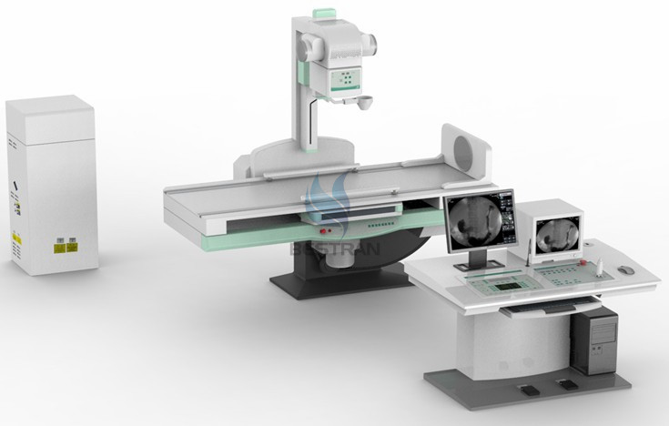
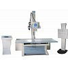
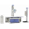
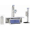
High Frequency Digital Fluoroscopy X-ray system (65KW, 800mA)
Application:
The unit is available for medical teaching, research, medical units for gastrointestinal X-ray spot film photography, remote control compartment, fluoroscopy and other Traumatic Radiography etc of X-ray routine examination, Can be used for gastrointestinal imaging, cholangiography, Urinary tract imaging and Deep vein imaging and other imagings ; Can used for fluoroscopy of esophagus, breast, gastrointestine, Abdomen and limbs and spot film photography; Can do the operation of Fracture reduction and Taking foreign body from in vivo .
Features :
1. Diagnostic table
·1)Visual display of man-machine interface, touch screen control, makes man-machine conversation more intuitive, convenient and easy to understand.
·2)Programmable controller technology, high control precision, good stability, excellent anti-jamming performance
·3)Inverter control technology, When the table move, it can soft start, soft stop, smooth no noise, more accurate positioning
·4)Reliable, high precision of angular displacement sensor technology, low dynamic noise, and long mechanical life.
·5)High-resolution of rotary encoders, high accuracy of auto-slice, security and stability.
·6)Rotation range of 90 ° ~ 0 ° ~ -25 °, can take routine examination of digestive organs , can also be used for a variety of special examination
·7)Humanized Design of diagnostic table lateral switch , near station positioning make the operation more convenient.
·8)Toshiba's high-quality image intensifier, to further enhance image sharpness, contrast, more easily, to clearly identify Lesions.
·9)Move a wide range of spot film device, apply the operation fo People do not move with the operation of motor way easily to complete taking from the throat, esophagus,To the lower abdomen of a series of examinations .
·10)Spot film box open on their own, safe and reliable operation.
2. Generator
•1)With X-ray Radiography and perspective function of generator, maximum output power of 65 kW.
•2)Output voltage of 150 kV.
•3)With a smaller, lighter, modular design.
•4)In the exposure process, kV and mA can be adjusted to achieve a constant radiation output.
•5)Large-screen LCD flat panel used to display the APR conventional conditions and practical projects.
•6)User-friendly system configuration.
•7)Operator can modify the APR technology items.
•8)Provide APR /-ray tube data downloads.
•9)A variety of automated diagnostic procedures, and operators with prompts
.•10)With serial RS232 communication interface
•11)Operation can be individually programmed for the APR, and APR and manual techniques for programming options
•12)Programmable settings, calibration, and conduct regular APR (by connecting an external computer).
•13)For ABS (Automatic Brightness Stabilization) circuit select four kinds of kV and mA curve.
Specifications:
|
Item |
Content |
Technical parameter |
|
|
Power supply |
Voltage |
380V±38V |
|
|
Frequency |
50Hz±1Hz |
||
|
Capacity |
≥85kVA |
||
|
internal resistor |
≤0.13Ω |
||
|
Digital X-ray high voltage system (import high-frequency generator )
|
|
Power Output |
65KW |
|
|
Inverter Frequency |
200kHz |
|
|
photography |
Tube Voltage |
40kv—150kv step regulation |
|
|
Tube Current |
10mA—800mA step regulation |
||
|
Exposure time |
1.0s—6300ms step regulation |
||
|
Control interface |
Touched LCD |
||
|
fluoroscopy |
Tube Voltage |
40kv—125kv step:2kv continuous regulation |
|
|
Tube Current |
0.5mA—6mA continuous regulation |
||
|
Automatic brightness for fluoroscopy IBS |
Automatic brightness tracking,multiple settings beforehand |
||
|
Digital Controlled X-ray Tube (Toshiba, Japanese) |
Model |
E7252 |
|
|
Tube Focus:Large Focus/Small Focus |
1.2mm /0.6mm |
||
|
Input Power |
Large focus:75kW small focus: 27kW |
||
|
thermal capacity |
210KJ |
||
|
Rotary anode speed |
2700rpm |
||
|
Micro-computer control digital tube remote diagnostic table
|
Material of the tabletop |
high strength、low absorb carbon fiber |
|
|
Rotation of the table |
90°~0°~-25° |
||
|
Transverse travel of table |
±110mm |
||
|
Longitudinal travel of the photography holder and film spot device |
≥720mm |
||
|
X-ray Focus—film gauge |
1100 mm—1500 mm |
||
|
Rotating foot plate |
±360° rotation |
||
|
Full film photography size |
8″×10″—14″×17″ |
||
|
Dividing method for spot film |
Full film, half dividing, three parts dividing, four parts dividing |
||
|
Control method for diagnostic table |
Remote、table control、frequency conversion soft starting and stopping |
||
|
beam limiter |
Electric Multi-leaf |
||
|
Voice interaction |
Two-way microphone system |
||
|
fixed bucky device for table |
Grid density: 103L/INCH, Grid ratio: 10:1,focusing distance: 120cm,fixed type: 15″×18″ |
||
|
Digital Image TV System |
Image Intensifier |
TOSHIBA integrated image intensifier with 400,000 pixels CCD camera (non-digital) / TOSHIBA 9'' image intensifier and 1 mega pixels CCD camera (digital) |
|
|
Monitor |
17〞medical high definition monitor, horizontal central resolution ratio :1000 lines,fringe: 800 lines,video bandwidth: 12.5MHz,50bit,image number/second: 25 frame |
||
|
Digital Picture Processing System (CCU Sentinel) |
High definition line by line output mode, 8 level noise reduction,store 8 image;LIH(freeze the last frame),the image can be turned vertically and horizontally,positive and negative image; OSD(monitor show) |
||


